Custom Labeled Liposome Service for In Vivo Imaging
Inquiry
Recently, there has been significant interest in imaging liposomes, and this nanoparticle-based approach holds promise for facilitating a combined therapeutic and diagnostic strategy. CD Formulation supports develop diverse liposome-based labeling products for in vivo imaging, utilizing cutting-edge technology and expertise.
What are Labeled Liposomes for In Vivo Imaging?
With the diverse design and application of liposomes, therapeutic diagnostic nanomedicine or nanotherapeutic diagnostics has emerged as a promising personalized medicine strategy. Labeled liposomes for in vivo imaging including radiolabelled liposomes can be utilized to investigate the in vivo biological distribution of liposomal delivery drugs and are also crucial for other therapeutic approaches such as radiation therapy and anti-angiogenic drugs for cancer patients. The combination of liposome labeling, and in vivo imaging techniques offers distinctive capabilities and innovative approaches for nanodiagnostic medicine, facilitating real-time visualization, in vivo monitoring, assessment, and personalized treatment.
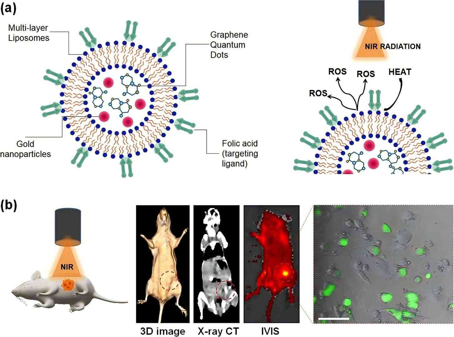 Fig.1 In vivo imaging of folic acid labeled liposomes. (Prasad, R., et al., 2020)
Our Custom Labeled Liposome Service for In Vivo Imaging
Fluorescently labeled liposomes are employed for in vivo imaging of target organs and inflamed sites. Our services are designed to support clients in high-quality liposomes for medical diagnostics and in vivo research.
We employ a variety of methodologies to label lipids with radioactive isotopes, resulting in the production of highly efficient liposome products that exhibit superior imaging performance, exceptional stability, and remarkable multifunctionality as carriers of radioactive isotopes for imaging and diagnostic applications.
Ultrasound-switchable fluorescence (USF) technology is an advanced high-resolution imaging technique recently developed. Our platform for ultrasound-switchable fluorescence imaging reagents can assist clients in creating various fluorescent groups to achieve high-resolution fluorescence imaging at the centimeter scale in deep tissues.
We can incorporate a variety of cutting-edge technologies (such as liposome targeting, nuclear magnetic imaging, near-infrared imaging, and ultrasonic imaging) to support clients in achieving specific imaging objectives.
Our Platforms and Techniques
| Techniques and Platforms |
Specifics |
| Fluorescent Labeling Technique |
- Offering a selection of fluorescent dyes suitable for in vivo imaging.
- Providing quantum dots for multiplexed labeling and enhanced detection sensitivity.
|
| Ultrasonic Switchable Fluorescence Imaging Near-infrared Liposome |
- Our platform for ultrasound-switchable fluorescence imaging reagents.
- Creating various fluorescent groups to achieve high-resolution fluorescence imaging at the centimeter scale in deep tissues.
|
| Radiolabeled Liposome Techniques |
- Supports different radioisotopes such as 18F, 64Cu, etc.).
- Supporting different radiolabeling methods including passive encapsulation, membrane labeling, and remote labeling-ionophore.
|
Why Choose CD Formulation?
- Techniques for labeling liposomes. We provide customized liposome probes and a diverse range of fluorescent molecules to support multi-channel flow cytometry experiments, as well as offer quantum dots for multi-color labeling options.
- The proficient teams. The technical team comprises seasoned and highly skilled experts proficient in liposome labeling technology for flow cytometry, with diverse multidisciplinary backgrounds and extensive field knowledge.
- Our high specificity labeling liposomes. Custom labeling ensures precise visualization of liposome-cell interactions.
Published Data
Technology: liposome-based in vivo tumor diagnosis technique
Journal: Commun Biol
IF: 6.5
Published: 2020
Results: The author developed a light-mediated photo-triggered strategy to enhance nanohybrid tumor accumulation. Additionally, doxorubicin hydrochloride was encapsulated in liposomes to demonstrate photo-triggered chemotherapy and functionalized with folate-targeting ligands. The encapsulated agents exhibited dual-modal imaging for in vivo tumor diagnosis due to their high contrast and emission characteristics. Targeted liposomes demonstrated tumor shrinkage mediated by near-infrared light (750 nm) due to the heat and ROS generated. Furthermore, compared to liposomes loaded with GQDs, which have been confirmed by anti-tumor studies, the liposomes showed excellent ROS scavenging ability. Therefore, this method and engineering system can pave the way for new directions in targeted imaging and cancer therapy.
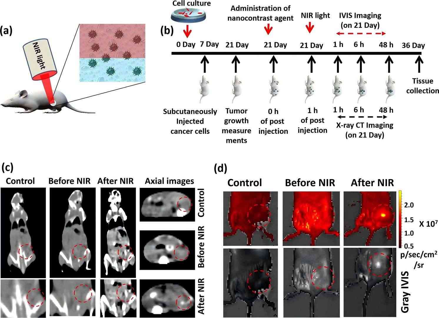 Fig.2 Images of near-infrared in vivo imaging system. (Prasad, R., et al., 2020)
As a leading company in nanoparticle development, CD Formulation is dedicated to providing excellent labeling of liposome products for in vivo imaging. Please do not hesitate to contact us if you require any assistance.
References
-
Prasad, R., Jain, N.K., et al. Liposomal nanotheranostics for multimode targeted in vivo bioimaging and near‐infrared light mediated cancer therapy. Commun Biol. 2020. 3. 284.
How It Works
STEP 2
We'll email you to provide your quote and confirm order details if applicable.
STEP 3
Execute the project with real-time communication, and deliver the final report promptly.
Related Services
