Custom Fluorescently Labeled Liposome Service
Inquiry
Fluorescent imaging is a vital tool in biomedical research, providing high sensitivity, resolution, and real-time imaging capabilities. CD Formulation is committed to advancing liposome technology for in vivo imaging focusing on producing stable fluorescent-labeled liposomes.
Why are Fluorescently Labeled Liposomes Needed?
Bioimaging has evolved into a highly dependable and sophisticated tool for diagnosing diverse diseases. Over recent decades, substantial advancements in imaging technology have facilitated the successful application of various advanced imaging methods in clinical diagnosis, thereby significantly enhancing human healthcare. As an advanced carrier system, liposomes can be loaded with fluorescent dyes enabling real-time tracking of their distribution and localization within the organism—a critical aspect when investigating targeted delivery to specific tissues or cells. Compared to alternative imaging techniques, fluorescent labeling offers a superior signal-to-noise ratio that contributes to obtaining clearer and more precise images of liposomes within intricate biological settings. Ultimately, fluorescently labeled liposomes enhance our capacity to investigate and comprehend their behavior within living organisms—thus aiding in the improved design and optimization of liposomal formulations across numerous biomedical applications.
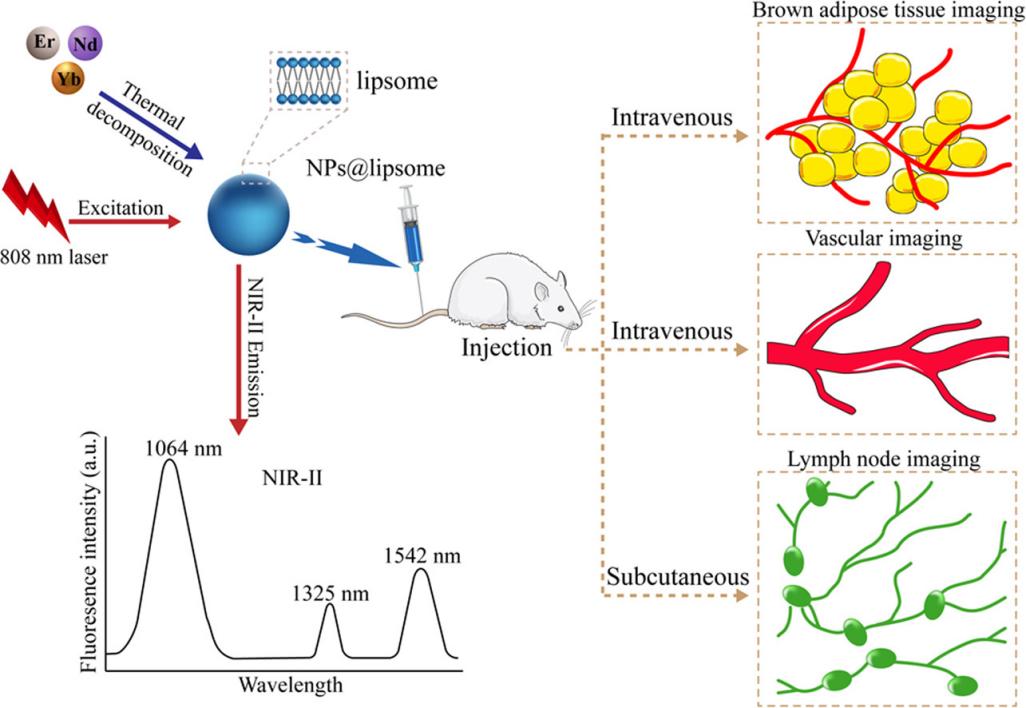 Fig.1 In vivo multifunctional fluorescence imaging using liposome-coated lanthanide nanoparticles. (Cheung CCL, et al., 2020)
Our Custom Fluorescently Labeled Liposome Service
Custom Fluorescent Probe Synthesis
We offer synthesis of a wide range of fluorescent probes suitable for IR-II imaging, including inorganic nanoparticles (carbon nanotubes, quantum dots, rare-earth-doped nanoparticles), organic molecules (e.g., conjugated polymers), and small molecule dyes. These liposome probes are integral for biomedical applications, especially in preclinical IR-II fluorescence imaging.
Custom Low Molecular Weight Fluorophores
Low molecular weight fluorescent dyes (with a typical molecular weight of ≈1,500 Da for near-infrared fluorophore groups) are the most commonly used tracers due to their well-defined chemical structure, tailored optical and chemical properties, and widespread commercial availability. Our available dyes include popular organic fluorescent group families such as rhodamine, BODIPY, indolocarbazole, porphyrin, and phthalocyanine.
Fluorescently Labeled Liposome Targeting
By combining fluorescent tracers with targeting components, the positioning and binding effects of the dye in the imaging area can be enhanced, as well as its pharmacokinetics. The targeting component may include antibodies, proteins or peptides, oligonucleotides, sugars, or other molecular templates known for their specific affinity to cellular compartments, cell receptors, biological fluids, or tissues. Utilizing liposomes as a novel strategy to alter the distribution of low molecular weight fluorescent groups in biological systems allows for encapsulation, adsorption, or grafting of fluorescent dyes onto surfaces.
Our Capabilities for Customizing Fluorescently Labeled Liposomes
| Techniques & Platforms |
Details |
| Fluorescent-labeled Liposome Probe Technology |
- To synthesize a series of fluorescent probes for IR-II imaging of various diseases, including inorganic nanoparticles (such as carbon nanotubes), quantum dots, rare earth-doped nanoparticles (RENPs), organic molecules (such as conjugated polymers) and small molecule dyes.
|
| Targeting Technology |
- Fluorescent tracer technology for binding to target components. The target components include antibodies, proteins or peptides, oligonucleotides, sugars, or other molecular templates known to have specific affinity for cellular compartments, cell receptors, biological fluids, or tissues.
|
| Low Molecular Weight Fluorophore Technology |
- Custom organic dyes such as rhodamine, BODIPY, and porphyrin.
|
Why Partner with CD Formulation?
- Advanced Labeling Techniques: Our precise labeling technology integrates fluorescent dyes with cell-specific molecules, ensuring accurate and targeted delivery for in vivo imaging.
- Cutting-edge Analysis Platforms: We provide advanced imaging and analysis services to study pharmacokinetics, biodistribution, and cellular uptake of fluorescently labeled liposomes, aiding preclinical research.
- Expert Team: Our team comprises specialists in molecular biology, chemistry, and pharmacy, with years of experience in R&D, guaranteeing high-quality service and innovative solutions.
Published Data
Technology: The liposome labeling technology by cancer-fighting drugs with fluorescent properties.
Journal: Sensors
IF: 3.4
Published: 2016
Results: This study aimed to develop a nanodevice for targeting anchored liposomes (with and without cholesterol) and encapsulating anti-cancer drugs and antisense N-myc gene oligonucleotides on their surface. Liposomes encapsulating adriamycin, rosetree, and etoposide were prepared to achieve this objective. Their fluorescence intensity was measured for further characterization, with an estimated encapsulation rate of 16%. The hybridization process of oligonucleotides forming nanostructures was investigated using spectrophotometry and electrochemical methods. It was determined that the concentrations of rosaline, adriamycin, and etoposide attached to the nanostructures in the gold nanoparticle-modified liposomes were 14 µg·mL−1, 5 µg·mL−1, and 2 µg·mL−1 respectively. These studies successfully demonstrated that liposomes are suitable for transporting anticancer drugs and antisense oligonucleotides while effectively inhibiting the expression of the N-myc gene.
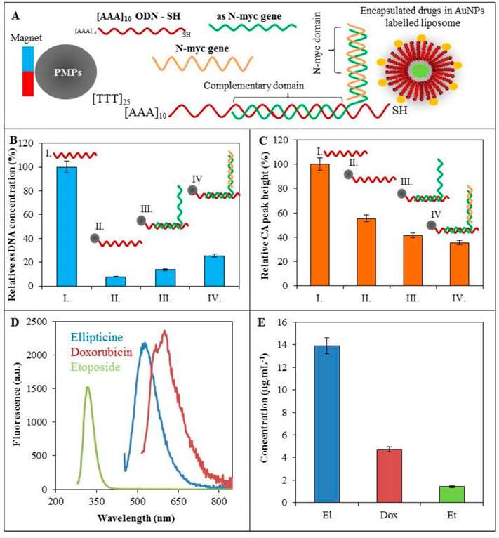 Fig.2 Schematic diagram showing the combination of AuNPs with fluorescently labeled modified liposomes. (Yang, J., et al., 2021)
As an expert in nanoparticles, CD Formulation is dedicated to delivering exceptional liposomes for in vivo imaging applications. Please do not hesitate to contact us if you require any assistance.
References
- Skalickova S, Nejdl L, et al. Fluorescence Characterization of Gold Modified Liposomes with Antisense N-myc DNA Bound to the Magnetisable Particles with Encapsulated Anticancer Drugs (Doxorubicin, Ellipticine and Etoposide). Sensors. 2016; 16(3):290.
- Yang, J., He, S., et al. In vivo multifunctional fluorescence imaging using liposome-coated lanthanide nanoparticles in near-infrared-II/IIa/IIb windows. Nano Today. 2021. 38, p.101120.
How It Works
STEP 2
We'll email you to provide your quote and confirm order details if applicable.
STEP 3
Execute the project with real-time communication, and deliver the final report promptly.
Related Services
