Nanoformulation for In Vivo Imaging Studies
Inquiry
As nanotechnology improves by leaps and bounds, nanoformulations not only come into wide use in disease treatment and drug delivery but also are widely used for in vivo imaging. CD Formulation attaches great significance to the development and production of nanoformulation for in vivo imaging studies and has accumulated rich research experience in many years of practice. We are able to provide customers with personalized customized solutions and nanoformulation customization services for in vivo imaging studies.
Why is Nanoformulation Important for In Vivo Imaging Studies?
Nanoformulations play an active role in in vivo imaging studies due to their ability to provide larger imaging payloads, higher sensitivity, multiplexing capabilities, and modular design. Using nanoformulation-based in vivo imaging techniques, we can observe deeply into the living body, which provides great opportunities for disease diagnosis and research. In the meantime, nanoformulations have become the key to producing high-resolution, high-contrast images and are widely used for precise disease diagnosis.
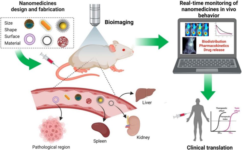
At the same time, due to the physical and chemical complexity of nanoformulations, we use advanced imaging techniques to visualize the metabolic behavior of systemically administered nanoformulations in situ. Thus, the use of in vivo imaging methods in the evaluation of nanoformulations can significantly improve the efficiency of nanoformulation development, rapidly advancing clinical translation and commercial applications of nanoformulations.
 Fig.1 Non-invasive imaging modalities can provide real-time monitoring of NMs fate in vivo and facilitate their clinical translation. (Ruslan G. Tuguntaev, et al. 2022)
Explore Our Nanoformulation-based Imaging Techniques
We take advantage of our advanced nanoformulation-based imaging techniques to offer anatomical, physiological, or molecular information about the in vivo systems to assist disease diagnosis.
Magnetic Resonance Imaging (MRI)
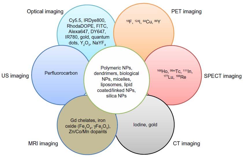
In the MRI imaging, we provide a variety of information about the body's interior, including anatomical, physiological, and even molecular information, utilizing our contrast agents (e.g., paramagnetic (Gd3+) and superparamagnetic (Fe3O4) nanoparticles).
Computed Tomography (CT) Imaging
Through CT scanners, we utilize radioactive X-rays multiple times to acquire signals from different angles and process them with computers to reconstruct three-dimensional images.
Positron Emission Tomography (PET) Imaging
We detect positron signals (gamma rays) emitted by radioactive isotopes or tracers by PET imaging to help with clinical diagnostics.
Single-photon Emission Computed Tomography (SPECT) Imaging
We detect different radioisotopes to visualize the system in the body by SPECT imaging to assist diagnostics.
Optical Imaging
In vivo optical imaging has the characteristics of non-invasive, highly sensitive, cost-effective, non-ionizing, and real-time imaging. Based on our professional knowledge and rich production experience, we fabricate plenty of best quality NIR fluorophores for in vivo imaging.
 Fig.2 Incorporation of various contrast agents for multimodality imaging. (Jaehong Key, et al. 2014)
Custom Versatile Nanomaterials for In Vivo Imaging Studies
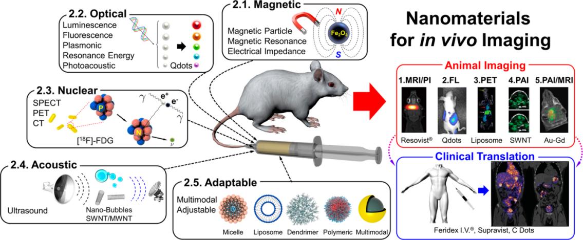
CD Formulation endeavors to gain a full understanding of the magnetic, optical, acoustic, and/or nuclear properties of nanomaterials and strives to pursue functionalized nanomaterial development.
Magnetic Nanomaterials
According to the requirements of in vivo imaging (such as using magnetic resonance imaging, magnetic particle imaging, magneto-motive imaging, and electrical impedance imaging), we customize magnetic nanomaterials based on Gd3+, Mn2+, and iron-based materials, such as gadolinium oxides (Gd2O3), gadolinium phosphates (GdPO4), GdF3:CeFn3 nanomaterials, spherical MnO nanomaterials, hollow mesoporous silica MnO nanomaterials (HMnO@mSiO2), iron oxide and iron alloy-based nanomaterials, conductive nanomaterials such as titania and gold, etc.
Optical Nanomaterials
Based on the performance of optical imaging, we design and produce fluorescent nanomaterials (such as quantum dots, qdots), carbon nanomaterials (such as carbon qdots), upconversion and persistent luminescent nanomaterials.
Nuclear Nanomaterials
We are keen on exploring and researching the fabrication of neclear nanomaterials for imaging, such as radioactive nanomaterials, chelated nanomaterials and intrinsically labeled nanomaterials.
Acoustic Nanomaterials
Our R&D center has also developed various nanobubbles, silica nanoparticles, and nanotubes for ultrasound imaging.
Adaptable Nanomaterials
To advance and optimize in vivo imaging techniques, we design and synthesize varieties of adaptable nanomaterials, including lipid nanoparticles, polymeric nanoparticles, liposomes, polymeric micelles, silicon-based nanomaterials, etc.
 Fig.3 Nanomaterials for in vivo imaging. (Bryan Ronain Smith, et al. 2017)
Custom Nanoformulation Development Services for In Vivo Imaging Studies
Nanoformulations for in vivo imaging studies can provide important information for disease diagnosis through various in vivo imaging methods. At our multiple nanotechnology platforms, CD Formulation is committed to designing and developing various nanomaterials for in vivo imaging, meeting customers' different needs, and promoting the rapid development of disease diagnostic technology.
Lipid Nanoparticle Development for Nanomedicine
CD Formulation also focuses on the development of lipid nanoparticles for in vivo imaging. We can customize personalized lipid nanoparticles for different in vivo imaging methods to aid the disease diagnosis or nanoformulation evaluation.
Liposome Development for Nanomedicine
CD Formulation specializes in the research of liposome development for in vivo imaging. At our liposome technology platform, we deeply design and optimize the liposome development process to fabricate best quality liposomes for in vivo imaging studies.
Polymeric Nanoparticle Development for Nanomedicine
CD Formulation is good at polymeric nanoparticle development for in vivo imaging. We are able to design and customize polymeric nanoparticles for in vivo imaging studies, rapidly promoting the progress of disease diagnostic technology.
Exosome Development for Nanomedicine
CD Formulation has been committed to developing exosomes for in vivo imaging for many years. And we have accumulated experimental experience in the exosome development process, we can offer personalized exosomes for in vivo imaging to support the disease diagnosis.
Why Choose CD Formulation?
- Hold versatile nanotechnology platforms for customizing class-quality nanoformulations.
- Have advanced nanomaterial-based in vivo imaging techniques to assist the disease diagnosis.
- Have extensive experience in nanoformulation evaluation.
Published Data
Technology: Novel nanomedicines based on advanced optical imaging technology
Journal: Advanced Drug Delivery Reviews
IF: 15.2
Published: 2024
Results:
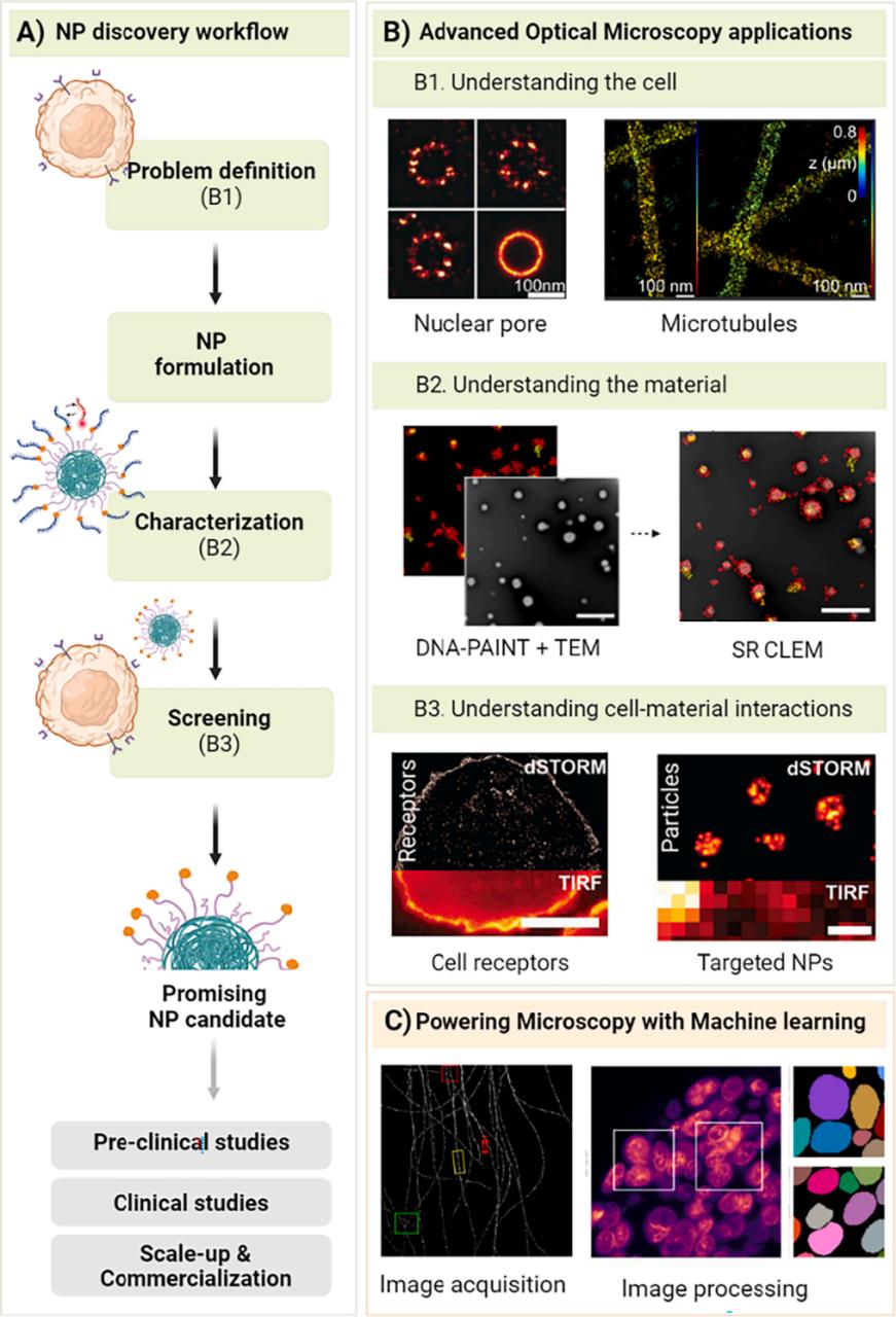
The authors use advanced optical imaging techniques for the rational design of nanomedicines and discuss in detail how to integrate advanced optical microscopy into different steps of the nanoparticle development workflow, namely by optimizing image acquisition, using machine learning (ML) to power microscopy, and image processing to help rationally design future nanomedicines. At the same time, the authors simplify the nanoparticle discovery workflow to find suitable or promising nanoparticle candidates for a given target diseased cell.
 Fig.4 Advanced optical imaging for the rational design of nanomedicines. (Ana Ortiz-Perez, et al. 2024)
In vivo imaging technology can not only assist in the rational design of nanoformulations but also has great application potential in disease diagnosis. CD Formulation has researched various nanoformulations for in vivo imaging studies, based on lipid nanoparticles, liposomes, polymeric nanoparticles, metal and inorganic nanoparticles, natural nanoparticles, etc. If you are interested in our nanoformulation for in vivo imaging studies, please contact us.
References
- Ruslan G. Tuguntaev, Abid Hussain, Chenxing Fu, et al. Bioimaging guided pharmaceutical evaluations of nanomedicines for clinical translations. Journal of Nanobiotechnology. 2022,20:236.
- Jaehong Key, James F Leary. Nanoparticles for multimodal in vivo imaging in nanomedicine. International Journal of Nanomedicine. 2014,9:711-726.
- Bryan Ronain Smith, Sanjiv Sam Gambhir. Nanomaterials for In Vivo Imaging. Chemical Reviews. 2017,117,901-986.
- Ana Ortiz-Perez, Miao Zhang, Laurence W Fitzpatrick, et al. Advanced optical imaging for the rational design of nanomedicines. Advanced Drug Delivery Reviews. 2024,2024,115138.
How It Works
STEP 2
We'll email you to provide your quote and confirm order details if applicable.
STEP 3
Execute the project with real-time communication, and deliver the final report promptly.
Related Services

 Fig.1 Non-invasive imaging modalities can provide real-time monitoring of NMs fate in vivo and facilitate their clinical translation. (Ruslan G. Tuguntaev, et al. 2022)
  Fig.2 Incorporation of various contrast agents for multimodality imaging. (Jaehong Key, et al. 2014)
  Fig.3 Nanomaterials for in vivo imaging. (Bryan Ronain Smith, et al. 2017)
  Fig.4 Advanced optical imaging for the rational design of nanomedicines. (Ana Ortiz-Perez, et al. 2024)
