Nanostructure Characteristics Analytical Service
Inquiry
Nanostructure characteristics of nanomedicines include the structure and shape of nanomedicines. The structure and shape of nanomedicines may affect the interaction of nanomedicines with proteins and cell membranes in the body, the release of drugs, and the degradation and transport of nanoparticles. CD Formulation can determine the structure and shape of nanomedicines through different technical methods such as electron microscopy to meet customers' different quality requirements.
Structures and Morphology of Nanomedicines
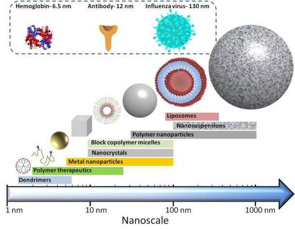
With our in-depth research into creative nanotechnology and nanoscience methods, we have successfully designed nanomaterials with different morphology, functions, and unique physical, chemical, and biological properties and applied them in nanomedicine development and manufacturing. Nanostructures prepared by different nanotechnologies include vesicles, solid nanoparticles, hollow nanoparticles, core-shell structures or multilayer structures, etc. And the common shapes of nanomedicines include spherical, spherical, rod-shaped, or fibrous shapes, etc.
 Fig.1 Different structures of nanomedicines and their approximate sizes. (Deepali Sharma, et al. 2020)
Why Conduct Nanostructure Characteristics Analysis?
Nanomedicines have scale effects based on nanostructures, and different nanomedicine structures and shapes may lead to differences in drug quality. The structural shape of nanomedicines can be detected by different technical methods such as electron microscopy. Therefore, when studying the structure and shape of nanomedicines, we can choose appropriate methods to detect and control the existence form and/or crystal state of the encapsulated drugs in nanomedicines to ensure the reliability of drug quality.
Our Nanostructure Characteristics Analytical Methods
CD Formulation is focused on nanotechnology to find out the solutions for nanomedicines and come up with low-cost, safe, and highly sensitive techniques for drug development and more efficient nanostructure characterization techniques. The nanostructure analytical methods we explore, and study include electron microscopy (EM), powder X-ray diffraction (PXRD), differential scanning calorimetry (DSC), polarized light microscopy, etc.
Electron Microscopy
Electron microscopy (EM) is a technique for obtaining high-resolution images of nanomedicine samples. There are two main types: transmission electron microscope (TEM) and scanning electron microscope (SEM). We can use this technology to study the structural characteristics of nanomedicines and provide researchers with detailed structural information about different nanostructures.
Powder X-ray Diffraction
The X-ray powder diffraction method is a fast, accurate, and efficient non-destructive testing technology for materials. We can use the X-ray powder diffraction method to characterize the crystal structure and its change rules. Through the Bragg equation, we can use X-rays of known wavelengths to solve for the crystal interplanar spacing to obtain crystal structure information.
Differential Scanning Calorimetry
Differential scanning calorimetry (DSC) is a thermal analysis method that can be used to determine a variety of thermodynamic and kinetic parameters, such as crystallization rate, polymer crystallinity, sample purity, etc.
Polarized Light Microscopy
Polarized light microscopy is a method of changing ordinary light into polarized light for microscopic examination to identify whether a substance is monorefractive (isotropic) or birefringent (anisotropic, a basic characteristic of crystals).
Our Workflow of Nanostructure Characteristics Analysis
Our team has lots of experience in nanostructure characteristics analysis and can quickly respond and work on your inquiry about nanostructure characteristics analysis, and then we can send you our final test report in just following four steps.
 Fig.2 Our workflow of nanostructure characteristics analysis. (CD Formulation)
Why Choose Us to Analyze Nanostructure Characteristics?
- Relying on our advantaged equipment, we can offer nanostructure characteristics analytical services using appropriate methods, such as EM, PXRD, DSC, etc.
- Our core team has rich experience in the characterization services of nanomedicines, including nanostructure characteristics analysis.
- We have complete analysis and testing service management specifications, a relatively fast sample circulation cycle, and can quickly respond to your detailed requirements for nanostructure characteristics analysis of nanomedicines.
Published Data
Technology: HR-TEM detection technology
Journal: Materials & Design
IF: 8.4
Published: 2023
Results:
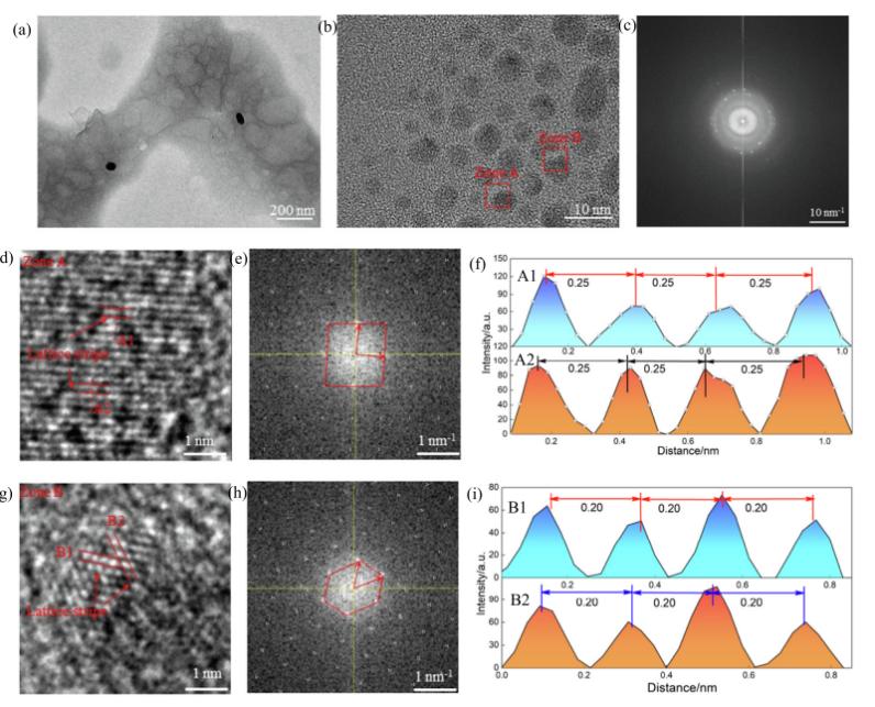
The authors used transmission electron microscopy (TEM), X-ray photoelectron spectroscopy (XPS), Fourier transform infrared (FTIR) spectrometer, and Raman spectrometer to conduct hydroxyl oxide graphene (HGO)@Ag-nanoparticle (Nps) coating. Analysis of physical and chemical properties. The authors used an electrochemical workstation to study the electrochemical performance of the HRO@Ag-Nps coating in 0.9% NaCl and 10% bovine serum albumin (BSA) solutions. The authors also used TEM to capture the composite nanostructure between the cysteine of the HRO@Ag-Nps coating and the methylene group of the BSA solution. The results of this study indicate that the HGO@Ag-Nps coating exhibits higher corrosion resistance. Here is the TEM measurement picture of the nanostructure of the HRO@Ag-Nps coating.
 Fig.3 Nanostructures with low-(a) and high-(b) magnifications, electron diffraction pattern (c), nanostructures (d, g), electron diffraction patterns (e, h) and atomic spacing (f, i) on zones of A and B. (Kong Weicheng, et al. 2023)
CD Formulation has a variety of world-class analytical equipment and a team with rich experience to provide nanostructure characteristics analytical services for your nanoformulation development needs. If you are interested in our services, please kindly contact us for your detailed information.
References
- Deepali Sharma, Chaudhery Mustansar Hussain. Smart nanomaterials in pharmaceutical analysis. Arabian Journal of Chemistry. 2020,13: 3319-3343.
- Kong Weicheng, Qi Yushi, Deng Yiming, et al. Nanostructural characteristic and electrochemical performance of hydroxy graphene oxide@Ag-nanoparticles coating: Combined TEM and DFT calculation. Materials & Design. 2023,229:111881.
How It Works
STEP 2
We'll email you to provide your quote and confirm order details if applicable.
STEP 3
Execute the project with real-time communication, and deliver the final report promptly.
Related Services

 Fig.1 Different structures of nanomedicines and their approximate sizes. (Deepali Sharma, et al. 2020)
 Fig.2 Our workflow of nanostructure characteristics analysis. (CD Formulation)
 Fig.3 Nanostructures with low-(a) and high-(b) magnifications, electron diffraction pattern (c), nanostructures (d, g), electron diffraction patterns (e, h) and atomic spacing (f, i) on zones of A and B. (Kong Weicheng, et al. 2023)
