Nanoformulation Cellular Uptake Assay
Inquiry
An essential aspect of the interaction between nanomaterials and cells involves the internalization of nanoparticles (NPs). Examining the mechanisms and pathways through which nanoparticles are absorbed into cells can offer significant insights into the impacts and potential hazards of nanomaterials on bodily tissues and organs. This comprehension is vital for ensuring the secure application of nanomaterials in the field of biomedicine. CD Formulation provides nanoformulation cellular uptake assay services, facilitating the investigation of cellular uptake pathways and experimental techniques associated with nanoparticles.
Cellular Uptake Mechanism and Pathway of Nanoparticles
Nanoparticles are artificial structures of nanoscale size that are created externally. They are engineered to transport particular molecules, such as genes, medications, and contrast agents, to specific locations within cells, such as the cytosol or nucleus. The manner in which nanoparticles are processed within cells is crucial for their effectiveness. Therefore, a thorough comprehension of how cells absorb and transport nanoparticles is vital for developing effective and safe nanomedicines. This involves adjusting the physical and chemical characteristics of nanoparticles to enhance their targeting, absorption, and transport within cells.
Phagocytosis
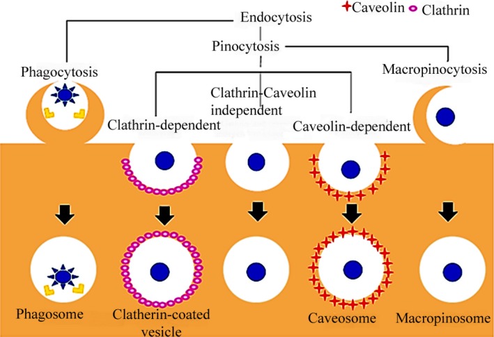
Phagocytosis is the process by which specialized cells within the immune system transport particles larger than 500 nm through a receptor-regulated mechanism. These particles are identified by small proteins such as immunoglobulins, complement proteins, and serum proteins. Upon recognition, the proteins coat the particles and bind to receptors on the cell membrane. This binding triggers a signaling cascade within the cell, leading to the extension of the cell membrane to engulf the particles. Subsequently, the engulfed particles are degraded by lysosomal enzymes, and the receptors are recycled to the cell membrane for further use.
Clathrin-mediated Endocytosis
Clathrin-mediated endocytosis serves as a primary cellular process for the uptake of nutrients and plasma membrane constituents, including cholesterol facilitated by low-density lipoprotein and iron transported by the transferrin carrier.
Caveolae-dependent Endocytosis
Caveolae-dependent endocytosis is a specialized form of cellular internalization, characterized by the formation of small, flask-shaped invaginations in the plasma membrane known as caveolae. These structures are rich in cholesterol and are stabilized by the protein caveolin. Caveolae mediate the uptake of certain molecules, including lipids and signaling molecules, without the need for clathrin, and they play a crucial role in various cellular processes such as signal transduction and the regulation of membrane composition.
Clathrin/caveolae Independent Endocytosis
Clathrin/caveolae-independent endocytosis occurs in cells lacking clathrin and caveolae. These cells take up different substances, such as cytosol, interleukin-2, and growth hormone, through alternative pathways that require specific lipid compositions (such as cholesterol) without clathrin and caveolae.
Macropinocytosis
Macropinocytosis is the actin-driven endocytosis of extracellular fluid-phase macromolecules. The activation of macropinocytosis is caused by the activation of tyrosine protease receptors, which leads to actin aggregation, and cell membrane ruffling to form large and irregular primitive endocytic vesicles and ultimately complete the uptake.
 Fig.1 Scheme for the main pathways of nanoparticle endocytosis. (Sara Salatin, et al. 2017)
Factors Affecting the Efficacy of Nanoparticle Uptake
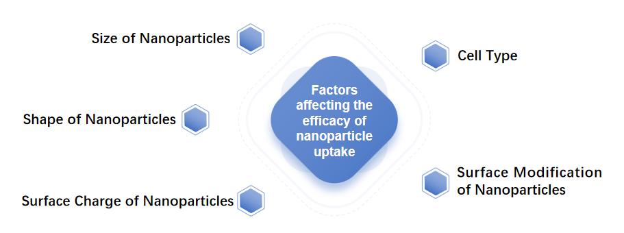
The uptake of nanoparticles is a complex process and is affected by multiple factors, such as particle concentration, particle size, particle morphology, particle surface modification, cell type, time, etc.
Particle Size: Cells can take up particles ranging from a few nanometers to hundreds of nanometers, and particle size is one of the important factors affecting cell uptake. Different cells have different optimal sizes for taking up different nanoparticles.
Shape: The shape of nanoparticles is also an important factor affecting cellular uptake.
Surface Charge: The surface charge of the nanoparticles also affects the interaction of the nanoparticles with the cell membrane. The surface of the cell membrane has a negative charge, so nanoparticles with a positive charge on the surface can more easily enter the cell.
Surface Modification of Nanoparticles: The surface of nanoparticles can be surface modified through some chemical ligands, such as antibodies, peptides, and sugars. The concentration, spatial distribution, and molecular weight of these chemical ligands will affect the cellular uptake of nanoparticles.
Cell Type: Different cell types have different rates, amounts, and pathways of nanoparticle uptake.
 Fig.2 Factors affecting the efficacy of nanoparticle uptake. (CD Formulation)
Explore Our Nanoformulation Cellular Uptake Assay
Relying on our high-end instrumentation, CD Formulation has explored and developed multiple techniques for studying nanoformulation cellular uptake assay, such as. Transmission electron microscopy, scanning electron microscopy, atomic force microscopy, flow cytometry, etc.
- Super-Resolution Fluorescence Microscopy
- Transmission Electron Microscopy
- Atomic Force Microscopy
- Scanning Electron Microscopy
- Light-Scattering Microscopy
- Flow Cytometry
- Dark-Field Microscopy
- Photoacoustic Microscopy
- Surface-Enhanced Raman Scattering
- Laser Ablation ICP-MS
- Correlative Microscopy
- X-Ray Adsorption Near-Edge Spectroscopy
Why Choose Us for Nanoformulation Cellular Uptake Assay?
- With the help of our advanced instruments and equipment, our technical team has accumulated rich experience in constructing appropriate techniques of nanoformulation cellular uptake assay.
- We hold multiple instruments and equipment for nanoformulation cellular uptake assay, such as TEM, AFM, SEM, light-scattering microscopy, etc.
- We highly value your requirements for nanoformulation cellular uptake assay and are able to provide you with personalized research plans quickly and efficiently.
Published Data
Technology: Entropy-Mediated Nanoparticle Cellular Uptake
Journal: Small Science
IF: 12.7
Published: 2023
Results:
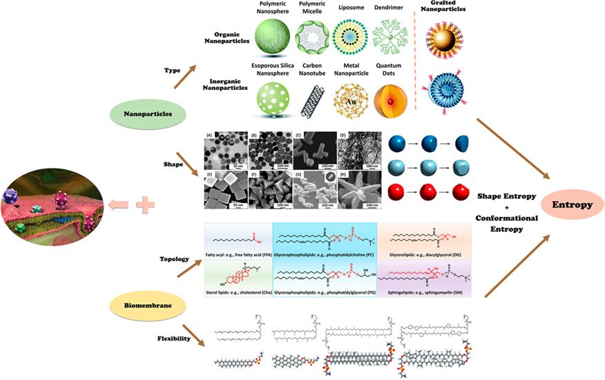
The authors introduce the main types of entropy in biological systems, the basic physical principles of entropy forces, and the entropy effects that occur at the interface between nanoparticles and cell membranes in different biological processes. The physics of entropy generation during cellular uptake is emphasized. The authors explain the physical mechanisms behind the structure and kinetics of some complex and/or unique phenomena observed during cellular uptake and promote the use of entropy control strategies in the design and development of new functional systems and materials to achieve beneficial biomedical applications.
 Fig.3 The schematic diagram of entropy-mediated NP cellular uptake. (Haixiao Wan, et al. 2023)
CD Formulation has accumulated a wealth of practical experience in the nanoformulation cellular uptake assay and can provide professional technical support to researchers in exploring the cellular interactions of nanoparticles. If you require our nanoformulation cellular uptake assay services, please feel free to contact us for detailed discussions.
References
- Sara Salatin, Ahmad Yari Khosroushahi. Overviews on the cellular uptake mechanism of polysaccharide colloidal nanoparticles. J Cell Mol Med. 2017, 21(9): 1668-1686.
- Haixiao Wan, Duo Xu, Lijuan Gao, et al. Entropy-Mediated Nanoparticle Cellular Uptake. Small Science. 2023,4,2300078.
How It Works
STEP 2
We'll email you to provide your quote and confirm order details if applicable.
STEP 3
Execute the project with real-time communication, and deliver the final report promptly.
Related Services

 Fig.1 Scheme for the main pathways of nanoparticle endocytosis. (Sara Salatin, et al. 2017)
 Fig.2 Factors affecting the efficacy of nanoparticle uptake. (CD Formulation)
 Fig.3 The schematic diagram of entropy-mediated NP cellular uptake. (Haixiao Wan, et al. 2023)
