Microscopic Morphology Testing of Nanoparticles
Inquiry
Nanoparticles come in many shapes and their names are derived from their different shapes. For example, there are spherical nanospheres, nanoreefs, nanoboxes, nanoclusters, nanotubes, etc. Controlling the morphology of nanoparticles is essential to exploit their properties for applications in a variety of emerging technologies. CD Formulation can offer microscopic morphology testing services of nanoformulations utilizing our world-class instruments and equipment such as TEM, SET, AFM, NMR, etc.
How to Describe the Morphology of Nanoparticles?
The morphological characteristics of nanoparticles to be taken into account are roughness, sphericity and aspect ratio.
- Roughness: Roughness represents the controllable deviation of a spherical nanoparticle from the ideal state of a uniform spherical object and can accurately describe the detailed properties of the nanoparticle. The roughness of nanoparticles has a significant impact on their properties and functionality, and adjusting the surface roughness of particles can simultaneously achieve the performance of all particles with different shapes, sizes and surface properties.
- Sphericity: Sphericity and Roundness are two different measures of morphological characteristics. Sphericity mainly depends on the elongation. It only considers whether the long and short axes of the particles are close to each other but does not consider the edges and corners. Roundness mainly depends on the sharpness of the object's angular protrusions (protrusions) and dents (dents).
- Aspect Ratio: High aspect ratio and low aspect ratio nanoparticles are classified separately. High aspect ratio nanoparticles include nanotubes and nanowires with various shapes, such as spirals, zigzags, ribbons, or possibly nanowires with diameters that vary with length. Small aspect ratio morphologies include spheres, ovals, cubes, prisms, spirals or cylinders. Aggregations of many particles exist in the form of powders, suspensions or colloids.
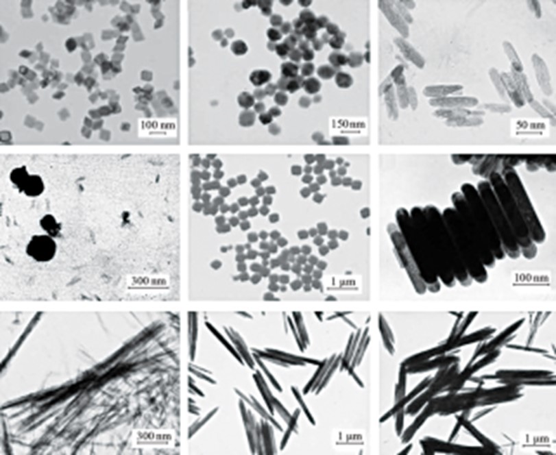 Fig.1 Nanoparticle morphology.
Why is Nanoparticle Morphology Important?
Nanoparticle morphology (size, shape, and composition) and surface chemistry are the determining factors underpinning the efficacy of nanomaterials in disease treatment applications. The size, shape, and surface chemistry of nanoparticles can strongly influence their critical properties, such as interactions with different biological fluids and interfaces, which affect the delivery of bioactive substances and modulate therapeutic properties. Nanoparticle morphology has been shown by scientists to significantly influence properties such as circulation time, cell interactions, and flow behavior.
Our Nanoparticle Morphology Testing Techniques
Common shapes of nanoformulations include spheres, spherical shapes, rods or fibers, etc. The structural shape of nanoformulations can be detected through different technical methods, such as electron microscopy. CD Formulation has explored and researched microscopic morphology testing techniques for the nanoproperty characterization of nanoformulations, such as brunauer-emmett-teller (BET) technique, transmission electron microscope (TEM), scanning electron microscopy (SEM), atomic force microscope (AFM), etc.
Brunauer-Emmett-Teller (BET) Technique
BET technology is based on the principle of physical adsorption of gases on solid surfaces, which we use to quantify the surface area, pore size and distribution, and accessible pore volume of a given nanomaterial with the help of nitrogen's adsorption and desorption isotherms.
Transmission Electron Microscope (TEM) Technique
One of the primary techniques we utilize is high-resolution transmission electron microscopy (HRTEM) as an effective imaging method for analyzing nanoscale substances. This tool allows us to observe the atomic-level details of nanomaterials, including their size, shape, uniformity, and lattice structure. HRTEM is frequently employed to assess the characteristics such as shape, size, morphology, layer thickness, crystallinity, defects, and interface structure of both synthesized and modified nanomaterials, as well as multi-component nanomaterials.
High-angle annular dark field scanning transmission electron microscopy (HAADF-STEM): We utilize this technique to provide "Z-contrast" images while using quantitative HAADF-STEM tomography and image analysis to obtain the morphology and internal structure of the nanoparticles.
Liquid Cell Transmission Electron Microscopy (LC-TEM): We use LC-TEM to conduct real-time studies of the fundamental processes of nanoparticle growth in solution.
Scanning Electron Microscopy (SEM)
SEM is an established technique in electron microscopy. We utilize it to examine the surface of nanoformulations, analyze nanoscale materials, study nanoparticle growth, and assess how ligands impact nanoparticle morphology.
Atomic Force Microscope (AFM)
The AFM force-distance (F-D) measurement monitors the deflection of the AFM cantilever as a function of distance as it interacts with a surface. We obtained this information to evaluate its possible correlation with properties such as surface functionalization and ligand shell stiffness.
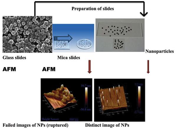 Fig.2 Nanoparticle observation using the AFM. (Syed Mahmood, et al. 2017)
Apart from the above techniques, we also offer such liking NMR and TGA for microscopy morphology testing of nanoformulations. Furthermore, spectroscopic techniques such as UV-visible spectrophotometry, small-angle X-ray scattering, and dynamic light scattering are also used to study the morphological characteristics of nanomaterials. These techniques are indirect methods based on physical or chemical properties of nanomaterials related to specific morphologies.
Why Choose Us for Nanoparticle Microscopic Morphology Testing?
- With the help of our advantaged equipment, we can provide microscopic morphology testing services using appropriate methods, such as BET technique, TEM, SEM, etc.
- Our technical team has been committed to the development and characterization of nanoformulations for years and has profound knowledge of the microscopic morphological testing of nanoformulations.
Published Data
Technology: Cryo-EM, SAXS and SANS techniques of SAM LNPs
Journal: ACS Nano
IF: 17.1
Published: 2024
Results:
The authors characterized the morphology of LNPs containing encapsulated SAM (SAM LNPs) and demonstrated that SAM changes its conformational structure when encapsulated in LNPs, becoming more compact than the free SAM form. A characteristic structure was observed in SAM LNPs, consisting of a lipid-rich core and an aqueous RNA-rich core, both surrounded by a DSPC-rich lipid shell. The authors used SANS and SAXS data to confirm that the prevailing morphology of LNPs consists of two core compartments with components unevenly distributed between the two cores and shell. Meanwhile, the authors constructed a capped cylindrical core-shell model with two internal compartments to capture the overall morphology of LNPs. These findings indicate that the bubble two-compartment structure can become a representative morphology among SAM LNPs, providing necessary support for the rational design of LNPs for mRNA delivery.
CD Formulation is a leading international nanoformulation development service company with many years of experience in microscopic morphological testing and can fully support your nanoformulation development and manufacturing needs. If you are interested in our microscopic morphological testing services, please contact us for in-depth communication.
References
- Syed Mahmood, Uttam K. Mandal. Morphological Characterization of Lipid Structured Nanoparticles by Atomic Force Microscopy while Minimizing the Formation of Failed Artefacts. Current Nanomaterials. 2017,2(1):24-32.
- Jacob L. Thelen, Wellington Leite, Volker S. Urban, et al. Morphological Characterization of Self-Amplifying mRNA Lipid Nanoparticles. ACS Nano. 2024,18, (2):1464-1476.
How It Works
STEP 2
We'll email you to provide your quote and confirm order details if applicable.
STEP 3
Execute the project with real-time communication, and deliver the final report promptly.
Related Services