Custom Labeled Liposome Service for Flow Cytometry
Inquiry
Integrating flow cytometry and labeling techniques offers a promising platform for disease diagnosis, treatment, and studies on various biological processes. CD Formulation is committed to the development of diverse liposome-based labeling products for flow cytometry, utilizing state-of-the-art technology and expertise.
What are Labeled Liposomes for Flow Cytometry?
Metabolic research has long been a pivotal focus within life sciences. Flow cytometry serves as a high-throughput and highly sensitive cell analysis technique that is progressively gaining widespread adoption in metabolic studies. It holds substantial relevance in areas such as infectious diseases, healthcare management, autoimmune disorders, and hematologic conditions including tumors and primary immunodeficiencies. Experiments utilizing flow cytometry techniques for tasks like cytokine detection and immune typing impose stringent requirements on diverse fluorescently labeled antibodies and fluorophore selection due to their direct impact on detection efficacy. Fluorescent-labeled liposome particles present a novel avenue for cellular metabolic investigations.
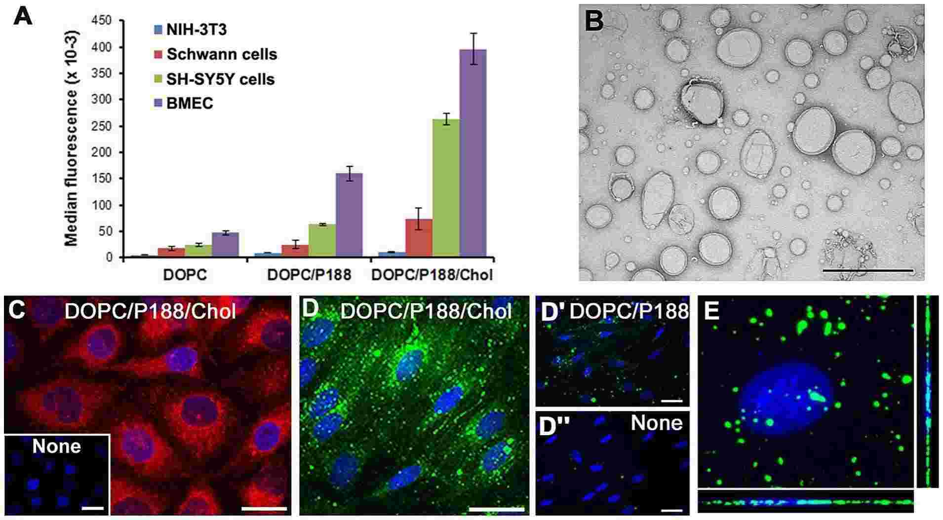 Fig.1 Quantitative assessment of the intracellular uptake of fluorescent liposomes. (Lee, Sooyeon, et al., 2013)
Custom Labeled Liposome Service for Flow Cytometry
The technology of flow cytometry facilitates rapid quantitative analysis and sorting of individual cells or other biological particles (such as microspheres, bacteria, and miniature model organisms) in a liquid stream. The utilization of liposomes in flow cytometry primarily encompasses cell transfection and liposome probes.
Liposomal formulations for cellular transfection
Liposomes serve as essential tools for in vivo and in vitro delivery, primarily utilized to encapsulate synthetic cationic lipids and DNA through fusion for cellular entry. Compared to alternative methods, liposomes for DNA delivery into diverse eukaryotic cells offer superior efficiency and reproducibility. We assist our clients in developing gene therapy products based on liposomes by providing fluorescently labeled liposome products.
Fluorescently labeled liposome customization
Fluorescein is categorized into four groups based on the excitation light wavelength, specifically 375nm, 405nm, 488nm, and 633nm. Among these, commonly used fluoresceins include those with excitation wavelengths of 488nm and 633nm, such as FITC, PE, APC, PI, etc. We are able to tailor liposome probes for flow cytometry to accommodate various fluorescence excitation wavelengths. We also provide quantum dots for multicolor labeling options, enabling multiplexed flow cytometry experiments.
Our Platforms and Techniques
| Techniques and Platforms |
Specifics |
| Fluorescent labeling technique |
- Fluorophores: Offering a selection of fluorescent dyes (e.g., FITC, PE, APC) or proteins (e.g., GFP, RFP) suitable for flow cytometry.
- Quantum Dots: Providing quantum dots for multiplexed labeling and enhanced detection sensitivity.
|
| Application exploration platform |
- Cellular Uptake Studies: Assessing liposome uptake and intracellular localization.
- Targeting Studies: Evaluating targeting efficiency to specific cell types or tissues.
- Drug Delivery: Monitoring drug release kinetics and efficacy.
|
Why Choose CD Formulation?
- Outstanding techniques for labeling liposomes. We provide customized liposome probes and a diverse range of fluorescent molecules to support multi-channel flow cytometry experiments, as well as offer quantum dots for multi-color labeling options.
- The proficient teams. The technical team comprises seasoned and highly skilled experts proficient in liposome labeling technology for flow cytometry, with diverse multidisciplinary backgrounds and extensive field knowledge.
- Our high specificity labeling liposomes. Custom labeling ensures precise visualization of liposome-cell interactions.
Published Data
Technology: fluorescently labeled liposome technique
Journal: Nanoscale
IF: 8.3
Published: 2018
Results: Liposome-targeted research necessitates the use of flow cytometry or microscopy. This aspect of the study relies on fluorescently labeling liposomes to ascertain their intracellular localization. The authors employed size exclusion chromatography (SEC) to quantitatively assess the propensity of commonly used fluorescent lipids to dissociate from liposomes during incubation in human plasma, revealing that some frequently utilized fluorescent lipid labels exhibited up to 75% dissociation, while others showed less than 10% dissociation. To evaluate the significance of this discovery, they utilized flow cytometry to measure the uptake of different fluorescently labeled liposomes by peripheral blood leukocytes and observed notable differences in uptake correlated with the trend of dissociation.
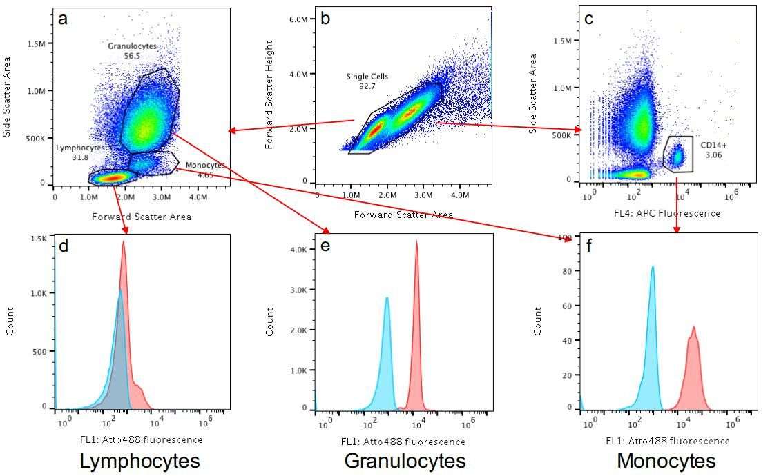 Fig.2 Gating strategy used to analyze flow cytometry data. (Münter R, et al., 2018)
As a leading company in nanoparticle development, CD Formulation is dedicated to providing excellent labeling of liposome products for flow cytometry. Please do not hesitate to contact us if you require any assistance.
References
-
Lee, Sooyeon; Tari, Ana; et al. Liposomes to Target Peripheral Neurons and Schwann Cells. PloS one. 2013, 8. e78724. 10.1371.
- Münter R, Kristensen K, et al. Dissociation of fluorescently labeled lipids from liposomes in biological environments challenges the interpretation of uptake studies. Nanoscale. 2018; 10(48): 22720-4.
How It Works
STEP 2
We'll email you to provide your quote and confirm order details if applicable.
STEP 3
Execute the project with real-time communication, and deliver the final report promptly.
Related Services
