Protein Content Determination
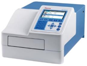
InquiryCD Formulation has detection platforms such as microplate reader (ELISA Reader) and UV-visible spectrophotometer, which can provide good protein content determination of biological preparations. At the same time, it provides the necessary reference basis for product packaging, specific activity calculation, limit control of residual impurities and determination of other physical and bio-pharmaceuticals.
Why to Perform Protein Content Determination?

Protein content determination is one of the most commonly used and basic analytical methods in biochemical research. With the continuous development of DNA recombinant technology, various target proteins can be obtained using different expression systems. However, the expression levels of proteins in different systems can be high or low, so it is extremely important to choose an appropriate method to measure the concentration of proteins.
Our Methods of Protein Content Determination
CD Formulation will develop personalized proposals to detect protein content based on the customer's needs. Currently, we can provide 6 methods for determination of protein content, namely Kjeldahl method, biuret method, Folin method (Lowry method), biuret method, 2,2'-biquinoline-4, 4'-dicarboxylic acid method (BCA method), Coomassie brilliant blue method (Bradford method) and UV-visible spectrophotometry, high-performance liquid chromatography (HPLC method) and enzyme-linked immunosorbent method (ELISA method).
Equipment: Kjeldahl nitrogen analyzer
Testing principle: This method is based on the fact that more than two peptide bonds contained in protein molecules form a purple-red complex with Cu2+ in an alkaline solution. It has maximum light absorption at 540nm, and its color depth is proportional to the protein concentration within a certain range. Use the protein reference solution as a standard curve and use colorimetric method to determine the protein content in the test product.
Equipment: UV-Vis Spectrophotometer
- Lowry method and BCA method
Testing principle: These methods are based on the peptide bond contained in the protein molecule chelating with Cu2+ in an alkaline solution to form a protein-copper complex (the first step of the biuret reaction). This complex reduces the phosphomolybdic acid of the phenolic reagent to produce a blue compound. At the same time, under alkaline conditions, the phenol reagent is easily reduced by tyrosine, tryptophan, and cysteine in the protein to produce a blue reaction. Within a certain range, the color depth is proportional to the protein concentration. Use the protein reference solution as a standard curve, and use colorimetric method to determine the protein content in the test sample.
Equipment: UV-Vis Spectrophotometer
Testing principle: This method belongs to the dye binding method. Coomassie Brilliant Blue G250 is a methyl-substituted triphenylmethane containing two sulfonic acid groups per molecule and is acidic. The sulfonic acid group can combine with basic amino acids (arginine) and aromatic amino acids in protein molecules through van der Waals forces to form a stable dye-protein blue complex. There is a maximum absorption peak at the wavelength of 595nm, and within a certain concentration range, the color depth of the complex is proportional to the protein content. Use the protein reference solution as a standard curve, and use colorimetric method to determine the protein content in the test sample.
Equipment: UV-Vis Spectrophotometer or microplate reader (ELISA Reader)
- UV-visible spectrophotometry
Testing principle: Aromatic amino acids in protein molecules, including tryptophan, lysine, and phenylalanine, contain conjugated double bonds that can absorb ultraviolet light. They have a maximum absorption peak at a wavelength of 280 nm, and the absorbance at this wavelength is related to its concentration. into a linear relationship. By detecting the wavelength of the target protein at 280nm with a spectrophotometer, the concentration of the target protein can be calculated according to the Beer-Lambert law.
Equipment: UV-Vis Spectrophotometer
Testing principle: When each component of the sample dissolved in the mobile phase passes through the stationary phase, due to the different size and strength of the interaction with the stationary phase (adsorption, distribution, exclusion, affinity), the residence time in the stationary phase is different, and thus flows out of the stationary phase one after another.
Equipment: HPLC
Testing principle: The specific protein content determination method is to immobilize a certain concentration of antigen or antibody in a microwell plate through physical adsorption and add it to the sample to be tested. The color intensity of the enzyme label indirectly reflects the presence or absence of the sample being tested or the amount.
Equipment: Microplate reader(ELISA Reader)
Our Workflow for Protein Content Determination
Here are following six steps to easily solve your requirements.

Advantages of Our Services
- Quickly assess and propose a professional testing plan based on your requirements for protein content determinationof bio-pharmaceuticals.
- To provide you with the most competitive market quotation and professional consulting services.
- Arrange the most efficient testing services for your project.
How to Contact Us?
If you have a requirement about our services, please contact us by phone or email, our colleagues will reply to you within three working days.
Related Services


