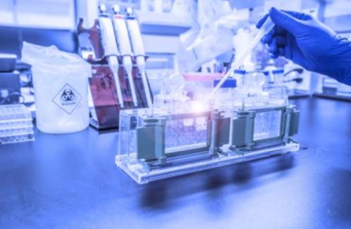
Exosome Characterization Services
Inquiry
In the study of exosomes, exosome identification is another key step after exosome separation and purification. Does the exosome you isolated have a typical exosome structure? Does it express specific exosome surface markers? Does the size fall within the conventional size range of exosomes? Exosome identification can provide answers to these questions. Exosome identification is to evaluate the presence, quantity and integrity of exosomes in enriched exosome samples through different experiments to evaluate the isolation and purification effect of exosomes by researchers. This is a necessary prerequisite for all future experiments.
CD Formulation has an advanced equipment platform and an experienced professional team to provide you with rigorous experimental design to ensure reliable analysis results and provide comprehensive support for your exosome identification.
Our Technology Platform for Exosomes Identification
Transmission Electron Microscopy (TEM)
Due to the small diameter of exosomes, which is generally between 30 and 150 nm, traditional optical microscopy cannot identify them. However, transmission electron microscopy can observe whether there are exosome-like structures (usually a saucer-shaped or hemispherical shape with a depression on one side) in the sample by gradually magnifying and imaging the sample after electrons emitted by an electron gun pass through it. It is a direct method for identifying exosomes and can also measure their particle size.
Nanoparticle Tracking Analysis (NTA)
Nanoparticle tracking analysis can track the motion of individual particles in a liquid, calculate the size of exosomes by analyzing the Brownian motion of the particles, and determine their concentration by counting. It is a commonly used method for exosome research. NTA technology is simple to handle and can quickly analyze the size distribution and concentration of exosomes, providing strong evidence for their existence.
Western blot (WB)
After the formation and release of exosomes, they will form their own unique marker proteins. Using Western blot to detect these marker proteins can identify exosomes.
Our Service
CD Formulation has rich experience in the identification of exosomes, and can provide comprehensive support for your exosome identification, including morphology, particle size and concentration, biomarker proteins and other aspects of identification. Specific methods include transmission electron microscopy (TEM), nanoparticle tracking analysis (NTA) and western blot (WB) identification. In addition, we can also provide a one-stop service for exosome extraction, identification, and subsequent functional experiments. With our extensive experience in exosomes, state-of-the-art equipment and dedicated team, you can rely on the exosome research service expertise of the CD Formulation. If you have relevant needs, please feel free to contact us! We look forward to working with you.
Advantages of Our Services
- Efficient service, short cycle: Experienced teams with cutting-edge domain expertise work efficiently, complete tasks and deliver results in a relatively short time.
- High-quality data: Accurate analysis and interpretation by a professional team.
- Personalized solutions: Provide customized solutions according to customer needs, and closely cooperate with research projects.
- Price concessions, perfect after-sales service: Reasonable and competitive prices, providing customers with comprehensive and professional after-sales services.
Related Services